


Osteoarthritis of the knee joint — progressing degenerative-degenerative pathology. Initially, it amazes cartilage tissue, and then in a destructive process to engage the bone structure.
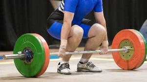
The causes of the development of arthrosis are increased physical load, low motor activity, metabolic and endocrine disorders. The head of the clinical manifestations of the disease — pain, increasing during walking, a functional failure of the knee replacement and its deformation. With the progression of the arthrosis develops stiffness (complete or partial immobilization of the completion of the joints). When placing a diagnosis of an orthopedist focuses on the results of instrumental research — arthroscopy, x-ray, CT, MRI. In the treatment of arthrosis 1 and 2, the degree of severity of the used pharmacological drugs, physical therapy, carried out by the physiotherapy and massage. When ineffectiveness of conservative therapy or the identification of pathology on the final stage of the patient preparing for the endoprosthesis of the knee joint.
The characteristic features of the disease
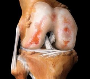
Osteoarthritis of the knee joint (gonarthrosis) is one of the most commonly diagnosed disease affecting this joint. Under the influence of external or internal negative factors in cartilage tissue arises from lack of nutrients. Consequently, it is violated trofika, slows down the regeneration of connective tissue structures. Happening premature aging of the hyaline cartilage. It narrows, crack, becomes rough, loses its firmness, flexibility and elasticity. Cartilage tissue can no longer perform its basic function — to reduce friction in the places of connection of the bones. Subchondral bone naked, paved, in them occur osteosclerotic changes. For stabilization of the knee joint during walking growth, flattened the edges of the bone plates with the formation of osteophytes (bone growths).
Osteophytes in the knee joint.
Primary articular arthrosis suffer from caught off guard initially healthy hyaline cartilage due to congenital reduction in their functional stamina. Secondary pathology occurs when they are already available defects of cartilage tissue. Can be triggered by previous injuries in the knee joints, the inflammatory process, aseptic necrosis, changes in the hormonal background disorders of metabolism.
| Also, the clinical-radiological stage of osteoarthritis of the knee joint | Specific symptoms |
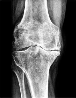
| The first | The mobility of the knee joint is slightly reduced, the contours of the joint space are becoming blurred, a little taper. On the edges of the bone plates to create a small amount of osteophytes |
| The second | During flexion or extension of the knee, is to hear the crunch, clicks, crackling sounds. Muscle slightly atrophy, significantly narrows the articular slit, forming a significant amount of osteophytes in bone tissue detectable subchondral osteosclerosis |
| The third | The knee joint is deformed, severely limiting his mobility. There was a complete or partial fusion of the articular slit, a large amount of bone growths, subchondral cysts, pathological formations, freely moving in the cavity of the completion of the joints |
Causes and precipitating factors
Impetus to the development of destructive-degenerative process in hyaline cartilage commonly becomes several negative factors. The cause of the development of arthrosis of the knee joint in the pediatric and adolescent age are disorders of the formation of the ligament-tendon apparatus, dysplasia of the. Again, due to hereditary predisposition. Trigger destruction of the cartilage can various injuries — fractures, severe contusions, sprains, a partial or complete break of the ligaments, muscles, tendons, meniscus. Excessive loading of the joint leads to the development of pathology. Post-traumatic arthritis develops after a few (3 to 5) years after the damage to the connective tissues or bone structures. Pathology can occur after surgery. And her cause becomes the inability of doctors and significant cartilage damage, and their slower regeneration. To the emergence of diseases also predispose to the following factors:
- being overweight, in which almost all structures of the musculoskeletal system, and especially the knee is subjected to excessive load;
- excessive physical activity often leading to microtraining cartilage tissue and further progression of the destructive processes;
- a sedentary lifestyle, which worsens the blood supply to the feet and formed a lack of nutrients in the hyaline skin;
- systemic inflammatory and degenerative-degenerative pathology — rheumatoid, psoriatic arthritis, gout, osteoporosis, lupus erythematosus.
The clinical picture
In most cases, arthrosis of the knee joint manifested by pain, dull pain after regular physical activity. The cause of its occurrence becomes irritating osteophytes on the in the vicinity of the soft tissue, venous stasis, intra-articular hypertension, muscle cramps. For arthrosis is a typical "starter" pain, occurs because of swelling the completion of the joints, or reactive synovitis. When a person a long time, is in a sitting position, and then stands up, then when the first steps he felt some pain. This creates the so-called "siege" the pain of a periodic nature. Knee joints are walking "stuck" due to the pinch points of the damaged cartilage tissue between the two surfaces of finished joints. Also for the arthrosis is characterized by next symptoms:
- crepitus, or crunching when flexion or extension of the knee, arising from the bias of bone structures relatively to each other on the background of the Antoniushof cartilage;
- stiffness, the severity of which falls on at least the seams of the joint space;
- spasms of the muscles located in the area of the knee joint, usually occur to reduce the pain;
- the deformation of the completion of the joints, triggered by destructive changes of the subchondral bone.
When osteoarthritis a person is difficult to climb stairs and go on long walks due to lingering soreness in the knee. During the pathology is very often complicated by synovitis — inflammation of synovial shells. Clinically it is manifested by the formation of a rounded flexible seal, hyperemia, severe edema, increase of body temperature to 37.1—38 °C, In the absence of medical intervention arthrosis complicates a spontaneous and hemarthrosis (bleeding into the cavity of the knee joint), complete or partial loss of mobility, on osteonecrosis of the patelofemorální condyle, the outer subluxation of the patella.
Diagnosis
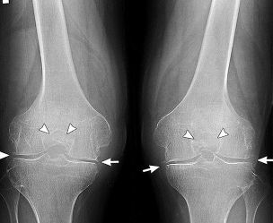
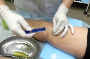
The clinical picture of arthrosis, especially complicated synovitis, quite similar with the symptoms of many inflammatory diseases of the musculoskeletal system. Therefore, performing differential diagnosis to exclude arthritis, inflammation of the tendons, of the tendovaginitis. Using instrumental research is defined by the condition of the knee completing the joints and the range of its functional activity. In the diagnosis of arthrosis the most informative radiography. On the images of the visible these body parts osteophytes, narrowed joint space, the deformation of bone structures (cysts, subchondral osteosclerosis). Symmetrical joint space narrowing of the knee joint are the joints affected in the articular arthrosis suffer. Likely to expose the operation will have to both limbs. A more detailed evaluation of changes in the hyaline cartilage allow ULTRASOUND, MRI, CT. Research is also carried out for the detection of inflammatory or degenerative lesion of the soft tissues, muscles, ligaments and tendons. In the case of materials is carried out by arthroscopy — a minimally invasive surgical manipulation. In the process of the implementation of the diagnostic procedures examined the inner surfaces of the completion of the joints, done the fence of biomaterials — synovial shells, synovial fluid, cartilage tissue. When synovitis by injection obtained by the pathological exudate on how to improve the well-being of the patient, as well as for examining its composition.
Tactics of treatment
Osteoarthritis of the knee 1 the degree of severity fairly well defies therapy using therapeutic physical training and longer-term use of chondroprotectors. Usually it is not necessary to use pain medication funds, as the symptoms expressed is not clear, or completely absent. When osteoarthritis of the knee joint 2 degrees also carried out a conservative treatment. But if the disease progresses, or is diagnosed with the destruction of significant quantities of cartilage tissue, then surgery is performed, usually the joint. In some cases, the patients indicated arthrodesis — artificial analogue of the stiffness, or complete immobilization of the knee joint.
Non-drug therapy
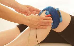
Sick joint arthrosis suffer from 2 or 3 degrees from the first days of treatment is recommended the wearing of rigid or semi-rigid brace, greatly limiting the mobility of the knee joint. When the small damage to the cartilage enough the use of soft elastic bandages — knee pads. Are fixed articulated arm, warn of more devastation of the tissues. Patients should reduce physical activity, do not lift heavy weights, avoid a long walk. They are appointed physiotherapy (5-10 visits), to improve blood circulation in the pain and stimulate the regeneration of connective tissue structures:
- magnetic therapy;
- laser;
- high-frequency electrotherapy;
- electrophoresis with a local anaesthetic and the cartilage of the knee;
- applique with ozokerite and (or) of paraffin.
To strengthen the muscular-ligamentous apparatus and to improve the motor function of the knee will help only the regular physical therapy exercises. Doctor of physical therapy picking up the exercises individually for each patient, with regard to the degree of arthrosis and physical preparation. The first exercises are held under his control. When osteoarthritis strictly prohibited any kind of intense movement. Improve blood circulation, but at the same time will trigger even greater mikrotemperaturnye cartilage. Movements should be smooth, measured, with a small amplitude. In the early stage of gonarthrosis incurred by destructive changes of the cartilage can be removed only with the help of medical physical education. Well established hirudotherapy or treatment of medical leeches. In the absence of measurements on the bottom are placed 3-4 leeches, usually in the area of the patella. Ringed worms bite the skin and inject into the blood saliva with a large number of biologically active substances.
Pharmacological preparations
For removal of severe pain in the knee are held drug blockade with corticosteroids. Hormonal remedies are usually combined with a local anesthetic. Intra-articular injection in osteoarthritis carried out only when the acute need, such as for corticosteroids are the characteristic expressed by the undesirable manifestations. Most often in the therapy of arthrosis applied non-steroidal anti-inflammatory preparations in various dosage forms:
- solutions for parenteral administration;
- pills;
- ointments and gels.
In the treatment scheme necessarily included chondroprotectors in the form of injections or tablets. These funds stimulate the regeneration of worn cartilage possess anti-inflammatory, analgesic and protiotokový effect. Every pain in the knee, should become a signal for immediate treatment to a podiatrist. On time carried out the treatment of arthrosis allow you to prevent the occurrence of serious complications, in some cases alert the patient's disability.