


Arthritis – is an inflammatory disease of the joints at which they are observed is destructive-degenerative processes, as is the defeat of the articular cartilage, ligaments, and bones, pathological changes in the synovial packaging (the membrane) and the joint capsule (in the shell of the joint, an airtight its closing and connecting to the bones in the joint). When osteoarthritis feel pain in the joint, possible restriction of mobility.
The causes of arthrosis
So far exactly is not known, what is the cause of the arthrosis, but there are a number of factors, aggravating the condition of the joint, changing its structure and functioning. The impetus for osteoarthritis can serve as:
- trauma (dislocation, the shoulder ligaments, fractures, contusions, penetrating wounds);
- static and dynamic loads associated with professional activities (in athletes, dancers, miners and other persons employed heavy physical exertion);
- obesity (under a constant load on the joint);
- hypothermia;
- infectious diseases (tuberculosis, chlamydia, influenza);
- violation of blood circulation;
- hormonal changes (pregnancy, menopause, menopause);
- metabolic disorders (osteoporosis (decreasing bone density), arthritis (inflammatory joint disease), gout (deposition of uric acid salts in the joints));
- autoimmune diseases (systemic lupus erythematosus (a disease in which the immune system recognises its own cells as foreign and fight with them), rheumatoid arthritis (the immune system mistakenly attacks a healthy joint));
- hemophilia a (due to hemophilia occur frequent bleeding into the joints);
- disorders of the endocrine system (thyroid disease);
- degenerative-degenerative changes (disease Pertusa (aseptic necrotizing process in the head of the hip joint), necrotizing process, which is part of the cartilage separates from the bone and passes into the joint cavity, causing necrosis of the spongy bones, which form a small fractures;
- congenital disorders (hip dysplasia of connective tissue (abnormal formation of tissue), hip dysplasia (abnormal development of the joint), congenital pathology of the upper and lower limbs);
- higher age;
- intoxication (an overdose of drugs, excessive alcohol drinking, drugs);
- delegated the operation of the joint;
- a sedentary lifestyle;
- heredity;
- beriberi.
The degree of arthrosis
The process of destruction of arthrosis is divided into 4 stages:
-
Zero degree of arthrosis – expressly referred to the morphological changes are not observed, but the allusions to disease, are already available. How is changing the composition of the joint fluid, therefore with the nutrition of the cartilage is deteriorating, therefore, its resistance to load decreases. In the morning after waking up helena after prolonged rest in a fixed state, it celebrates the difficulties in moving the joints, but during the day the stiffness is taking place. Such a condition is referred to as "starting pain". When the movement of the joint issued crisp, but the pain during movement is missing. For the first level is the typical pain, after a long, hard load on the joint, but the pain disappears immediately after a rest.
-
The first stage of osteoarthritis – the second stage is characterized by destruction of hyaline cartilage. Starting to morphological changes. X-ray shows the presence of newly formed osteophytes on the edges of the bone surface. The synovial capsule narrows. Celebrating joint space narrowing (narrowing of the distance between the capitals of the bones of the joint). Muscle function also exposed to negative influence. For the second stage arthrosis of the well-known constant pain, the crunching of the joints, sometimes claudication. The typical appearance of a "mechanical pain", generated during running, walking, exercise due to a reduction in depreciation of the ability of the cartilage.
-
The second degree of arthrosis – articular cartilage full-raddled, are large-scale slaughter. At this stage of the disease is dramatically evolving. It is characterized by a complete lack of synovial fluid, sharp pain, atrophy of muscles, shortening of the ligaments, destruction of the meniscus, the development of the jet synovitis (inflammation of the synovial membranes, the education of pus helena the accumulation of fluids, enlargement of the joint). The axis of the limbs deformed (X-figural and O-shaped curvature of the legs). Almost completely disappearing joint space, the deformed articular surface with the dimensions of the growth of osteophytes. Range of motion is limited, helena becomes impossible. It appears sensitivity to weather changes (humidity, low temperature, high atmospheric pressure). The third degree of arthrosis says about disability.
-
The third degree of osteoarthritis – on the fourth stage of the disease joint function lost forever. It will appear such a phenomenon as the sudden sharp pain in the affected joint, which does not soothe drugs. It can become stiffness (joining of bones), helena false joint, the unfolding is not a typical place for him. The fourth degree of osteoarthritis is indicative of the disability. Treatment involves only a quick intervention with an implant prosthesis.
Types of arthrosis
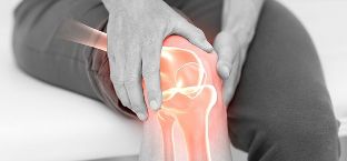
Osteoarthritis of the knee joint
Knee osteoarthritis is also called and deforming joint arthrosis suffer from the knee joint. The progression of the osteoarthritis brings pain and later causes the knee with his possible increase in volumes and an inflammatory result.
In athletes due to regular load on the knees, may develop the so-called osteoarthritis which attacks the knee joint, pain is felt in the front of the affected knee joint, the patella has a sharpened edge). For pain it is necessary to crunching and clicking of the joint.
Arthrosis of the hip joint
Also bears the name of coxarthrosis. This type of arthrosis hard drops and progresses faster other types of this disease, which sees to the disability. The defeat of one of the hip joint (one-sided osteoarthritis), may result in the defeat of the second (two-way osteoarthritis). Most likely available launched arthritis in the hip joints will cause the defeat of the knee joint and the spine. The observed lameness, shortening of the diseased limb, cracking, atrophy of the thigh muscles.
uncovertebral arthrosis
When uncovertebra osteoarthritis suffering from cervical joint.
In addition to the above factors, which have on the emergence of diseases of the joints, causes uncovertebra arthrosis can be the load on the cervical department, osteochondrosis (degenerative changes of the vertebrae, cartilage, intervertebral discs and adjacent tissue), the intervertebral hernia (a protrusion, a protrusion of the disc of the spine).
Osteoarthritis in cervical dysplasia accompanied by a reduction in cartilage, the seal and the deformation processes of the cervical, reduce the space, the growth of bone tissue in Boki due to narrowing of the space in the cervical region, compression of the nerve roots and narrowing of the spinal canal.
For uncovertebral arthrosis are typical manifestations of headache, sore throat, dizziness, vomiting, low sensitivity on the neck of helen's feelings chills in this area, an increase in blood pressure.
Osteoarthritis of the jaw joint (TMJ)
TMJ – temporal-mandibular joint. The joint is located closer to the ear, between the jaw and the temple on both sides of the skull. The cartilage of the jaw joint is not as strong as in other connections, so his defeat may causes pain. The disease is common in people aged 50 years old, suffering from mostly women. The result of arthrosis of the TMJ is difficulty in grinds food with the pain syndrome, impaired hearing, pain while speaking. In this case, the causes of osteoarthritis are: a change in the bite, congenital anomalies of the jaw, injuries to the jaw, stress, unfortunate dental treatment, the destroyed teeth, the older the age. The execution of the movement of the jaws, is published the click and crunch when you try to open the mouth, the jaw can move to the side.
Osteoarthritis of the elbow joint
This type of arthritis occurs less frequently other kinds of diseases, because the pressure on the ulnar joint is practically negligible. In the presence of inflammation of the place of localization of arthrosis swells and turns red, is present clicking, creaking, crunching during movement of the hand, a painful syndrome, slightly elevated body temperature.
Osteoarthritis of the feet
This type of arthritis also bears the name of coxarthrosis. Diseases of the ankle joint is characterized by increased fatigue when walking, swelling of (when you connect to inflammation in the process), pain syndrome (especially at night). Walk man, feeling the click in the ankle joint. In coxarthrosis damage relate to joints, muscles, tendons, nerves and blood vessels. Release of the tendons causes subluxation (the articular surface partially overlapping). When dislocation of the articular end are completely different.
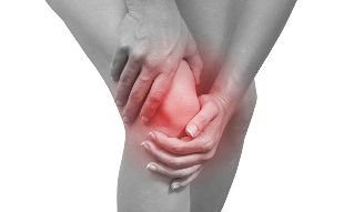
The symptoms of arthrosis
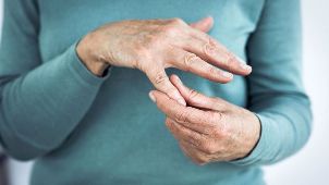
In the early stages of arthritis symptoms may not bother you, but on the authors of the disease is gaining speed, and showing the first and subsequent symptoms. The main symptom of osteoarthritis is pain depending on the site of the lesion (in the jaw and neck, on the shoulders, spine, elbows, wrists, fingers and toes, trunks, hip and ankle joint). The pains are dull in nature, but with the intensive progress of the disease worsen and become unbearable. The main symptoms of arthrosis:
- the initial pain (joint stiffness after sleep helena rest, passing in the course of the day);
- mechanical pain (arises after the work performance, load, running, exercises, walks);
- night pain (venous stagnation causes of pain in the middle of the night);
- weather dependent (weather changes lead to a worsening of the condition);
- crunching, creaking, cracking, helena unusual clicking in the joint;
- limited mobility;
- deformation of the affected joints;
- cramps, unpleasant muscle cramps;
- claudication;
- redness and swelling at the site of the affected joint (when using inflammation).
Diagnosis of arthrosis
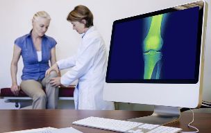
Self-diagnosis of "osteoarthritis" not to give. With annoy pain, crunching in the joint it is necessary to turn first to the general practitioner. It will assess both the patient and gives direction to the desired doctor, orthopedist, traumatologist, rheumatologist, surgeon helena neurologist.
Osteoarthritis diagnose the doctor-orthopedist. Before survey of the doctor collects the medical history (information about the patient, about the hereditary helena of chronic disease) and makes the control. Then the patient to send the survey.
Laboratory examination
In the diagnosis of arthrosis may be required the following tests:
- the general analysis of blood (blood from finger);
- biochemical analysis of blood (rheumatology test, the taking of blood from a vein);
- analysis of the urine;
- analysis of synovial fluid helena puncture of the affected joint (the plot through the introduction of needles into the joint).
Instrumental diagnosis
X-rays – is that a survey will reveal the stage of the arthrosis, even the slightest changes the structure of the joint and deformation using x-ray. Before the survey the patient should remove bijoux et, in order not to disrupt the result on the image. It is necessary to remove the clothing from that part of your body that will be examined (if you need an x-ray of the knee, it is necessary to take my pants off; the plaster is not excluded). Then the doctor says what position it should take on the image of the patient property. At the time of diagnosis the patient offered a lead apron groin of the guests and the guests of the thyroid gland, the apron prevents the passage at these locations x-ray radiation. Then take a picture.
The X-ray beam passes through soft tissue, but absorbed the hard (bones, teeth). So the soft cloth on the x-ray image shows dark colors and solid fabrics light. X-ray – the best method for the diagnosis of bone tissue, as it is in the pictures well displayed the bones and joints.
CT (computed tomography) – a method of beam diagnostics, which illustrate the body, the bones of the helena tissue in the cut, the layers. A CT scan is performed, if the x-ray is not enough informative and exactly osteoarthritis failed to detect. The procedure is carried out in the course of a few minutes. The patient lies down on a table, pre-clearing of clothing and metal ornaments. Then a ring of detectors (scanner) revolves around the reclining patient, and made the images. Lie in the CT examination should be motionless.
MRI (magnetic resonance imaging) – a method of radiological diagnosis. Differs from CT by a better distinction of soft tissue, but does not distinguish between the availability of calcium in the bones, unlike a CT scan, which examines in detail the bone tissue. MRI in osteoarthritis of the in the case, if you want to find out more information about the condition of the surrounding tissues helena to explore a key cause of the disease. Diagnosis using MRI expensive, but informative.
ULTRASOUND (ultrasound) – safe, not-invasive method, without radiation and harmful substances. The doctor has a sensor on the problem areas and the display shows the internal structure of the joint. This method of diagnosis of arthrosis of low-cost, safe and informative.
Weather
Arthritis has a good prognosis, if it goes again about the life of the patient, the disease is not fatal, but complications can lead to disability. However, to get from the arthrosis is not possible, because with age, the fractures occur faster, than the restoration of the damaged places. The disease itself progresses slowly and can develop for decades. The findings of the first signs of osteoarthritis can slow down and prevent further deformation and infringement of function of a joint with the help of treatment even in the initial stage.
Prevention
Often result in uneven, unnecessary exertion suffering from knee and hip joints, so it is necessary to calculate the power load and get rid of excess weight, if any, to get the joints working efficiently and cohesively.